Clay minerals are phyllosilicates that occur in very small sizes (“clay size”, corresponding to sizes less than 2 microns) and therefore cannot be identified under the petrographic microscope. The most usual analytical technique for its identification is X-Ray Diffractometry, which submits the samples to 3 analyzes (natural, glycolated and calcinated). The results, analyzed together, make it possible to identify the clay mineral(s) present.
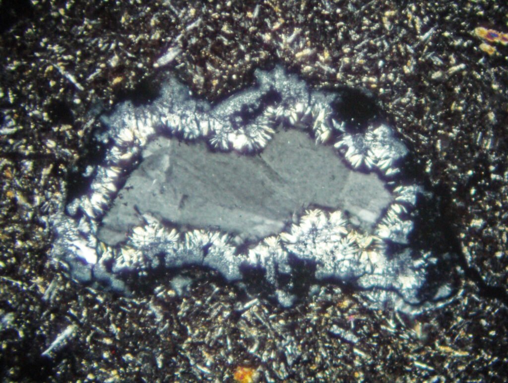
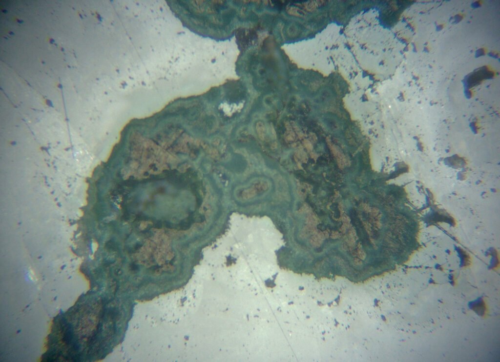
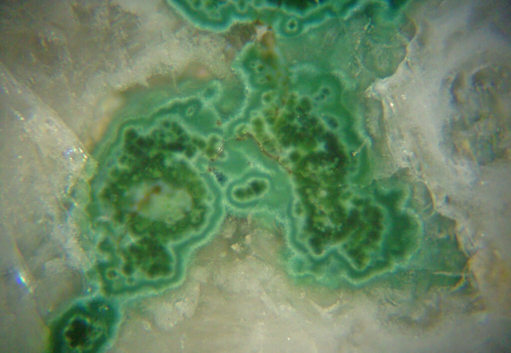
A very common clay mineral in thin sections is kaolinite, which is formed by alteration of feldspars (both potassic feldspars and plagioclase). The process is called “kaolinitization”. Not only kaolinite, but also clay minerals from the Montmorillonite Group are formed, generally by weathering processes.
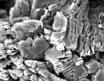
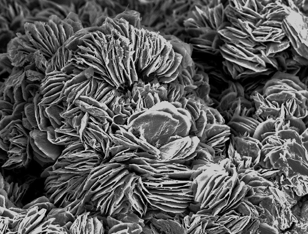
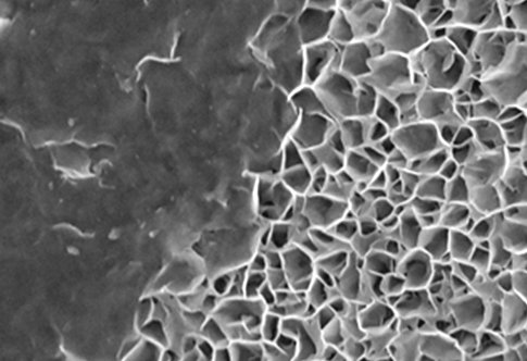
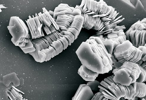
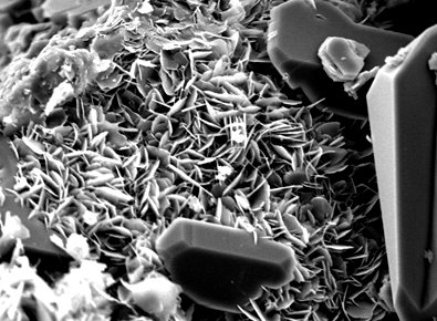
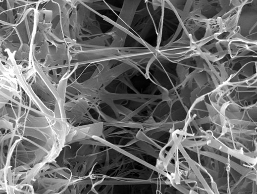
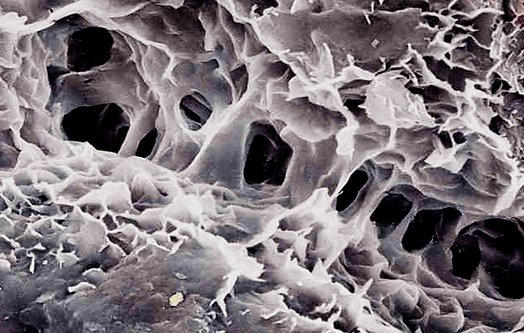
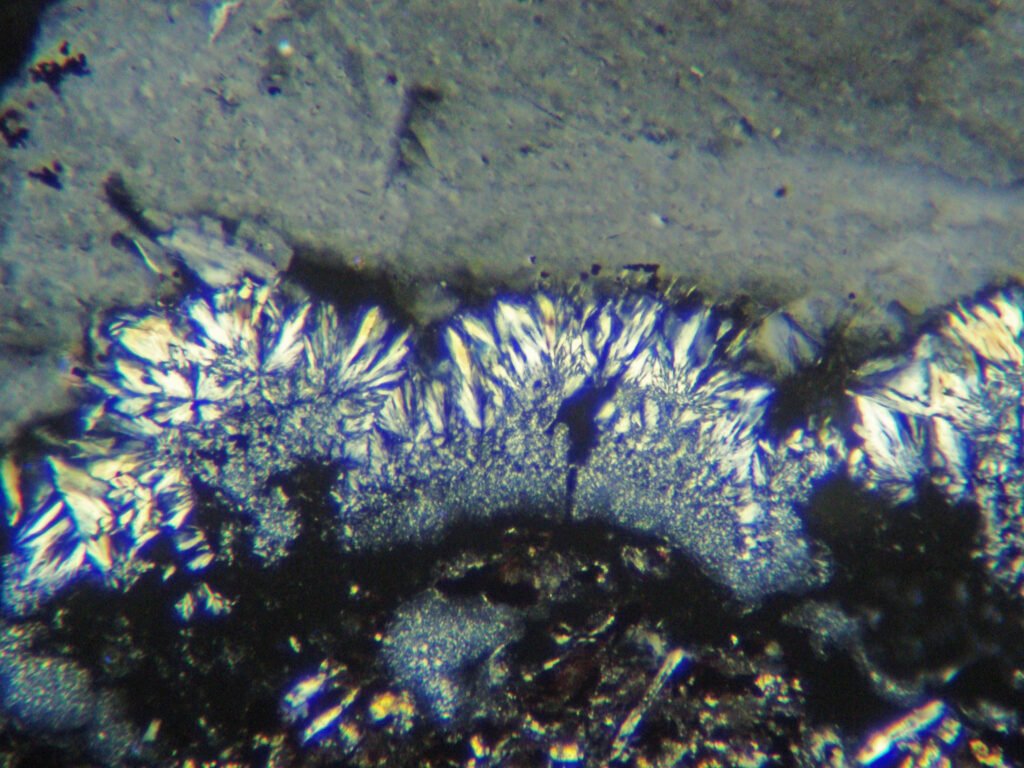
Clay minerals are phyllosilicates and, as such, develop crystals in the form of sheets or plates, which can form piles (“booklets”), fans, spherical aggregates, fibers, bridges, etc. Several clay minerals develop in rocks, such as smectites, kaolinite, dickite, illite and others.
We have to keep in mind, therefore, that in the thin sections and polished sections that we are analyzing there is, almost always, a large amount of clay minerals, of one or more species. Its optical identification is not possible, but its presence may indicate that the mineral alters easily, which sometimes can be diagnostic (feldspars alter, quartz not). The mention of clay minerals, referring to their appearance (colors) and the places where they develop (center or edge of the crystal, along fractures or cleavages, etc.) is important. The following are some Scanning Electron Microscope (SEM) images of clay minerals.